The Forearm Flexors
The forearm flexors form the muscle mass on the medial (inner) face of the forearm — the group that closes the wrist and flexes the fingers. When they develop trigger points, they produce the classic "golfer's elbow" pain (medial epicondylitis), but they can also cause tingling in the fingers and palm that faithfully mimics carpal tunnel syndrome (CTS) — even with a normal nerve conduction study.
The connection between forearm flexors and CTS symptoms is one of the most clinically relevant phenomena in myofascial medicine. Some patients operated on for carpal tunnel syndrome without postoperative improvement may have, among other possible causes, myofascial pseudo-CTS — symptoms identical to those of CTS, but caused by trigger points (TrPs) in the flexor digitorum superficialis, with normal electromyography and conduction velocity.
Myofascial Pseudo-CTS
TrPs in the flexor digitorum cause nocturnal paresthesias in the fingers and palm identical to CTS but with normal electroneuromyography — a critical diagnosis not to miss
Medial Epicondylitis
TrP1 of the proximal flexors is the muscular component of medial epicondylitis — combination of insertional tendinopathy + TrPs in the muscle belly
Typing and Technology
Prolonged keyboard, mouse, and smartphone use are the main modern drivers of flexor TrP development
Differential Diagnosis
Cubital tunnel syndrome (ulnar nerve), C8 radiculopathy, pronator teres syndrome, and true carpal tunnel must be systematically distinguished
Anatomy and Function
The main forearm flexors share a common origin at the medial epicondyle of the humerus — hence the name "golfer's elbow" for insertional tendinopathy at this site. The group includes the flexor carpi radialis, palmaris longus, flexor carpi ulnaris, flexor digitorum superficialis, and pronator teres.
Main Forearm Flexors
| Muscle | Main Function | Clinical Relevance |
|---|---|---|
| Flexor carpi radialis | Wrist flexion and radial deviation | TrPs cause pain in the radial wrist and base of the thumb |
| Palmaris longus | Wrist flexion (absent in 14%) | Less clinically important; used as a tendon graft |
| Flexor carpi ulnaris | Wrist flexion and ulnar deviation | TrPs cause pain in the pisiform and little finger |
| Flexor digitorum superficialis | Flexion of the middle phalanges (digits 2-5) | Main cause of pseudo-CTS; pressure in the carpal tunnel |
| Pronator teres | Forearm pronation | TrPs in pronator syndrome — proximal median nerve compression |

Trigger Points
Trigger points in the forearm flexors have two main patterns, with very different clinical implications: the proximal TrP, associated with medial epicondylitis, and the distal TrP, responsible for pseudo-CTS.
Referred Pain Pattern
The forearm flexors produce referred pain spanning the elbow to the fingers — a pattern that explains why these patients consult orthopedists (epicondylitis), neurologists (CTS, radiculopathy), and even rheumatologists (small-joint hand arthritis) before receiving the correct diagnosis.
Medial elbow pain when shaking hands or turning keys (TrP1 — medial epicondylitis)
Nocturnal finger tingling and numbness (TrP2 flexor digitorum — pseudo-CTS)
Palm and finger pain during long typing sessions
Difficulty opening jars, turning doorknobs, or gripping objects (weakness from TrPs)
Medial forearm pain radiating to the wrist (proximal TrP1)
Sensation of finger "stiffness" in the morning or after long typing sessions
Pain when carrying bags or gripping heavy objects (flexor contraction)
Paresthesias worse at night with the elbow flexed (distinguishing feature of cubital tunnel syndrome)
Causes and Risk Factors
The digital era has created a silent pandemic of forearm flexor TrPs. Activities that were once manual and alternating have become digital and static — typing, mouse use, and smartphones demand sustained low-intensity flexor contraction for hours, a pattern that favors TrP development more than intermittent intense effort.
Diagnosis
Diagnosis of forearm flexor TrPs is clinical. The key is the reproduction of pain or paresthesias by manual compression of the TrPs. For pseudo-CTS, the test reproducing paresthesias by the flexor digitorum TrP is the central diagnostic element — especially valuable when electroneuromyography is normal.
Clinical Evaluation of the Forearm Flexors
- Palpation of the medial epicondyle: local tenderness on compression (tendinous component of medial epicondylitis)
- Palpation of the proximal flexor belly: taut bands with tender nodules (TrP1)
- Palpation of the middle flexor digitorum belly: TrP2 that reproduces finger paresthesias — recognition sign
- Phalen's test: maximum wrist flexion for 60 sec — positive if finger paresthesias (true CTS)
- Tinel's sign at the carpal tunnel: percussion of the wrist — positive if paresthesias (true CTS)
- Grip strength test: bilateral dynamometry — weakness on the affected side
- Electroneuromyography: NORMAL in pseudo-CTS; altered (reduced conduction velocity) in true CTS
- Ultrasound of the common flexor tendon: thickening at the insertion (structural medial epicondylitis)
Differential Diagnosis
Forearm flexor symptoms overlap with important neurologic conditions. Distinguishing myofascial TrPs from nerve compression is decisive for treatment — especially before considering surgery.
Differential Diagnosis
Carpal Tunnel Syndrome (true)
Read more- Nocturnal paresthesias in the thumb, index, and middle fingers
- Positive Phalen and Tinel
- Thenar atrophy in advanced cases
Cubital Tunnel Syndrome (ulnar nerve)
- Paresthesias in the little finger and ulnar half of the ring finger
- Worsens with the elbow flexed
- Weakness of the intrinsic muscles (Froment's sign)
C8 Cervical Radiculopathy
Read more- Cervical pain + arm radiation
- Paresthesias in the little and ring fingers
- Grip weakness with a clear neurologic sign
Pronator Teres Syndrome
- Diffuse proximal forearm pain throughout the day (worse with activity)
- Worsens with resisted pronation
- Median nerve paresthesias WITHOUT nocturnal worsening (different from CTS)
Medial Epicondyle Fracture (trauma)
- Direct or indirect trauma to the elbow
- Localized edema
- Valgus instability of the elbow in young people
Pseudo-CTS versus true CTS: the diagnosis that changes treatment
Distinguishing true CTS from myofascial pseudo-CTS is one of the most important differential diagnoses in contemporary musculoskeletal medicine — with direct impact on the surgical decision. True CTS involves structural median nerve compression in the carpal tunnel, with documentable electroneuromyography changes: reduced sensory and motor conduction velocity of the median nerve across the wrist segment.
In myofascial pseudo-CTS, TrPs in the flexor digitorum superficialis raise carpal tunnel tension through musculotendinous taut bands without structural nerve compression — electroneuromyography is completely normal. Diagnosis is made by reproduction of paresthesias by manual compression of the TrP in the flexor digitorum belly. Operating on a patient with pseudo-CTS results in temporary improvement or no improvement at all, because the muscular cause remains intact.
True CTS
- True CTS involves structural median nerve compression in the carpal tunnel.
- Electroneuromyography shows reduced sensory and motor conduction velocity of the median nerve across the wrist segment.
- Phalen and Tinel tests are positive.
- The diagnosis is made by electroneuromyography.
Myofascial pseudo-CTS
- TrPs in the flexor digitorum superficialis raise carpal tunnel tension through musculotendinous taut bands, without structural nerve compression.
- Electroneuromyography is completely normal.
- Phalen and Tinel tests are negative.
- The diagnosis is made by reproduction of paresthesias by manual compression of the TrP in the flexor digitorum belly.
Cubital tunnel syndrome: when symptoms are in the little finger
Cubital tunnel syndrome is ulnar nerve compression at the elbow — the second most common upper-limb entrapment neuropathy. Unlike CTS (which affects the thumb, index, and middle finger via the median nerve), cubital tunnel causes paresthesias in the little finger and ulnar half of the ring finger. Worsening of paresthesias with elbow flexion (such as when talking on the phone or sleeping with the elbow bent) is highly suggestive of cubital tunnel. Tinel's sign over the ulnar nerve in the epitrochlear groove is the most specific clinical finding.
TrPs in the flexor carpi ulnaris and intrinsic hand muscles can produce pain in the ulnar forearm and hand, but without the clear pattern of worsening with elbow flexion. Electroneuromyography of the ulnar nerve at the elbow confirms or rules out cubital tunnel.
C8 radiculopathy: when the origin is in the cervical spine
Radiculopathy from C8 nerve root compression (spondylotic or from a C7-T1 disc herniation) produces pain and paresthesias in the little and ring fingers, possibly radiating along the ulnar forearm — a pattern similar to cubital tunnel, but with a cervical component. The clinically important distinction is cervical pain or radiation that starts in the neck and descends through the arm, plus neurologic findings such as a reduced triceps reflex and weakness of C8-innervated muscles, such as the flexor digitorum profundus and the intrinsic hand muscles. Cervical MRI and electroneuromyography confirm the diagnosis. Forearm flexor TrPs can coexist with C8 radiculopathy — myofascial and radicular pain overlap is common.
Treatments
Treating forearm flexor TrPs requires combining local treatment with ergonomic modification — without ergonomics, TrPs systematically recur because the same use patterns that caused them keep perpetuating them.
Diagnostic Confirmation
Electroneuromyography to investigate true CTS and C8 radiculopathy. Flexor tendon ultrasound if medial epicondylitis is suspected. Clinical evaluation of TrPs. Defining the myofascial versus structural contribution.
Local Treatment (0-4 weeks)
Needling of proximal and distal flexor TrPs. Acupuncture PC-3, PC-6, HT-3, LI-4. Local heat (TDP) for muscle relaxation. Stopping or modifying perpetuating activities.
Ergonomics (continuous)
Workstation evaluation: monitor height, keyboard position, ergonomic mouse. Wrist support at rest. 5-minute breaks every 45 minutes of typing. Flexor stretches during breaks.
Maintenance (after resolution)
Eccentric strengthening exercise program for medial tendinopathy. Gradual return to sport (racket, golf). Periodic ergonomic review. Return to physician if recurrence.
Myth vs. Fact
If electroneuromyography is normal, it isn't carpal tunnel syndrome and needs no treatment.
Normal electroneuromyography makes moderate or severe structural CTS less likely but does not rule it out in mild cases, and it does not rule out myofascial pseudo-CTS — which requires different treatment (needling of flexor TrPs). Telling the patient "normal" and dismissing them is an error that leaves a treatable cause undiagnosed.
Golfer's elbow is exclusively a tendon problem — it is enough to treat the insertion.
Medial epicondylitis combines insertional tendinopathy with active TrPs in the flexor belly. Treating only the insertion (local injections, shockwave) without deactivating the TrPs in the muscle belly produces partial improvement and frequent recurrence. Complete treatment includes the muscle-belly TrPs.
Do
- Workstation evaluation: monitor height, keyboard position, ergonomic mouse.
- 5-minute breaks every 45 minutes of typing.
- Flexor stretches during breaks.
Avoid
- Holding the mouse with a "forced grip" (stronger grip than necessary).
- Holding and typing on a smartphone with the wrist extended and fingers flexed — one of the most injurious flexor positions.
- Carrying bags and backpacks by a hand-held strap, especially with the arm extended.
- Treating forearm flexor TrPs requires combining local treatment with ergonomic modification.
- Without ergonomics, TrPs systematically recur because the same use patterns that caused them keep perpetuating them.
- After resolution, maintenance includes an eccentric strengthening exercise program for medial tendinopathy and gradual return to sport.
Acupuncture and Needling
Medical acupuncture for the forearm flexors mainly uses points on the pericardium (PC) and heart (HT) meridians, which run along the medial face of the forearm — a course that anatomically coincides with the forearm flexors. The addition of point LI-4 (Hegu) as a distal point enhances the analgesic effect.
For pseudo-CTS, the combination of needling the TrP in the flexor digitorum with PC-3-PC-6 electroacupuncture is a reasonable conservative option — the clinical rationale is that, with no structural compression of the median nerve (normal electroneuromyography), the myofascial cause may respond to local treatment. In mild to moderate cases of confirmed CTS, acupuncture is a conservative option before considering surgery, and the decision should be individualized by the physician. Usual clinical protocols involve cycles of 8-12 sessions, twice a week — numbers vary by study and individual response.
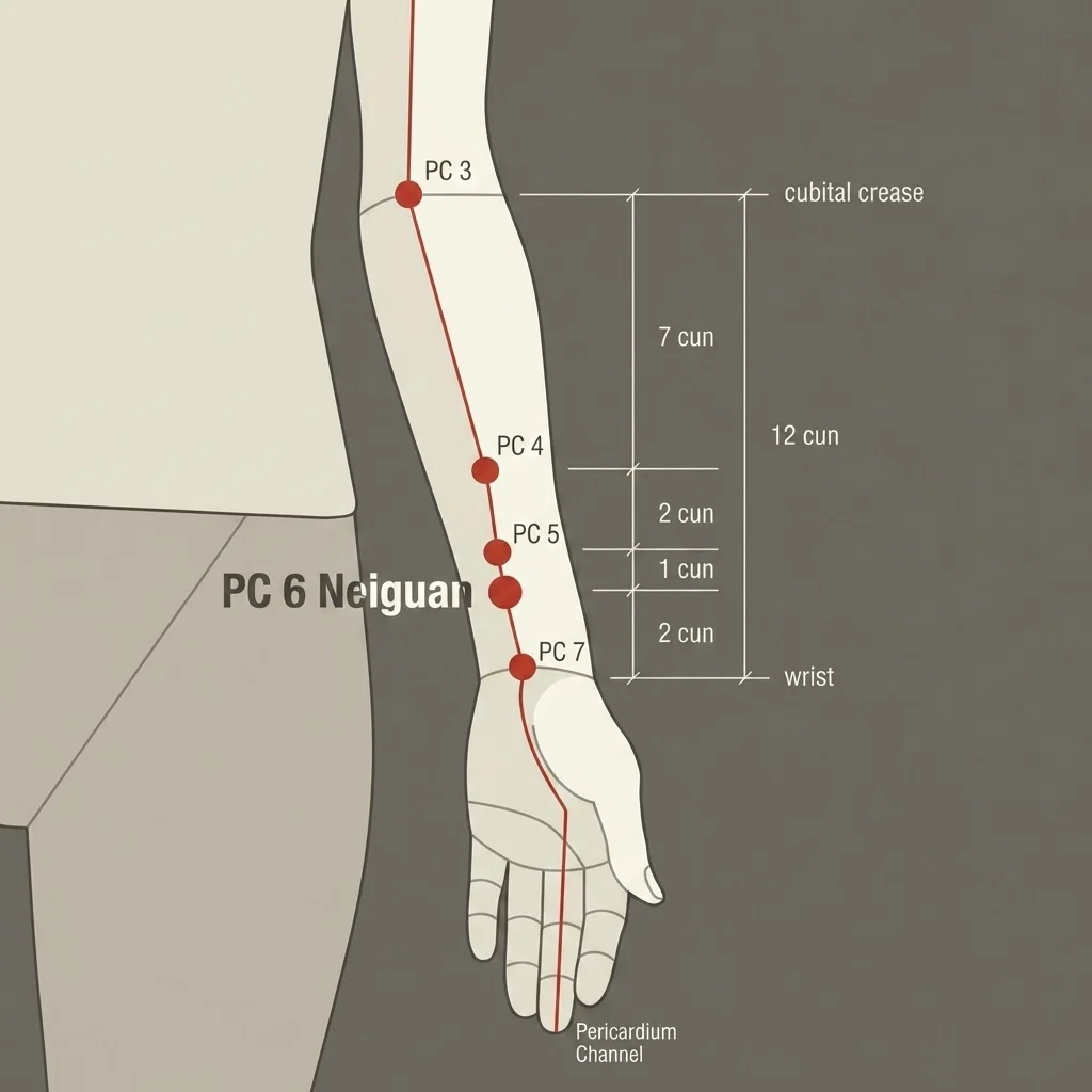
2 cun above the wrist, between the flexor tendons.
Prognosis
Forearm flexor TrPs have an excellent prognosis when ergonomic factors are corrected. Without ergonomic correction, recurrence is the rule — the muscle is again overloaded by the same activities that created the TrPs. With combined treatment (needling + ergonomics), resolution within 6-10 weeks is expected.
When to Seek Medical Care
Frequently Asked Questions
Forearm Flexors: Common Questions
Pseudo-carpal tunnel syndrome is a condition in which trigger points in the flexor digitorum superficialis cause symptoms identical to true carpal tunnel syndrome — nocturnal finger tingling, wrist and palm pain, numbness — but with completely normal electroneuromyography, since there is no structural median nerve compression. Diagnosis is made by reproducing the paresthesias through manual compression of the TrP in the flexor digitorum belly. The proposed treatment is TrP needling, after true CTS has been investigated, with evidence that is still limited.
Electroneuromyography is the key test: in true CTS, it shows reduced median nerve conduction velocity through the carpal tunnel; in myofascial pseudo-CTS, it is completely normal. Phalen (maximum wrist flexion for 60 seconds) and Tinel (wrist percussion) tests are positive in true CTS and negative in pseudo-CTS. When in doubt, the physician can compress the flexor digitorum TrP — if this reproduces the paresthesias, it confirms a myofascial contribution.
Medial epicondylitis is an insertional tendinopathy of the forearm flexors at the medial epicondyle of the humerus. It causes local pain at the inner elbow, aggravated by handshakes, turning keys, and loaded wrist flexion movements. It is called "golfer's elbow" because the golf swing is a typical mechanism, but it affects far more office workers, typists, and intensive smartphone users than golfers. Treatment includes the muscular component (flexor TrPs) and the tendinous component.
The relationship between typing and CTS is more complex than it appears. Typing itself, in correct posture with a neutral wrist, does not significantly increase the risk of true CTS. However, typing with the wrist forced into extension or flexion, with pressure on the carpal region, favors flexor TrP development (pseudo-CTS) and can contribute to true CTS in predisposed individuals. Keyboard position — which should keep the wrist straight — chair height, and regular breaks are decisive for prevention.
In myofascial pseudo-CTS (normal electroneuromyography), medical acupuncture with flexor TrP needling can significantly relieve symptoms without surgery. In mild to moderate true CTS, acupuncture is a conservative option before considering surgery — some studies suggest short- and medium-term benefit, with limited to moderate evidence strength, and the decision should be individualized by the physician. In severe CTS with thenar atrophy, surgery is necessary and acupuncture can complement postoperative rehabilitation.
Treatment of medial epicondylitis (golfer's elbow) combines an approach to the insertional tendinopathy and the TrPs in the muscle belly. For tendinopathy: eccentric flexor exercises (adapted Alfredson protocol), extracorporeal shockwave in chronic cases, and ultrasound-guided local injection if refractory. For TrPs: direct needling of the proximal flexor TrPs, acupuncture PC-3 and HT-3. Partial sports rest in the first 4 to 6 weeks. The use of a counterforce brace can relieve pain in the acute phase.
Before recommending surgery for CTS, at least 3 to 6 months of conservative treatment is advised, including a nighttime wrist splint in neutral position, ergonomic correction, medical acupuncture, and, if necessary, local corticosteroid injection. The exception is severe CTS with progressive thenar atrophy — in these cases surgery is urgent to prevent irreversible neurologic injury. Investigating myofascial pseudo-CTS is essential before considering surgery in cases with normal or borderline electroneuromyography.
Yes. Prolonged smartphone use is one of the main modern causes of flexor TrPs. The typical posture — holding the device with one hand and thumb-typing with the wrist extended and fingers flexed — overloads the flexors statically and repetitively. Average daily smartphone use commonly exceeds 4 hours globally — enough time to develop TrPs in predisposed individuals. Prevention: support the phone (table, stand), alternate hands, take breaks, and use voice commands when possible.
Yes, significantly. Active TrPs in the forearm flexors inhibit maximum muscle contraction force, which may reduce dynamometer-measured grip strength compared with the unaffected side. In grip-dependent sports (tennis, golf, volleyball, jiu-jitsu, climbing), this reduction directly impacts performance. After TrP treatment, grip strength may recover, in some cases within the first needling sessions.
Yes. Cervical radiculopathy, especially of the C8 root (compression at C7-T1), can cause tingling in the little and ring fingers, sometimes reaching the middle finger. Distinction from carpal tunnel relies on location (C8 affects ulnar fingers; CTS affects radial fingers), the presence of cervical pain radiating down the arm, and findings on cervical MRI. In patients with finger tingling and associated cervicalgia, investigating the cervical spine before opting for local wrist treatment is essential.