What Is Abdominal Diastasis?
Abdominal diastasis — technically termed diastasis of the rectus abdominis muscles (DRAM) — is the separation of the two bellies of the rectus abdominis muscle along the linea alba, the band of connective tissue that joins the recti at the midline of the abdomen. It is not a muscle tear, but a widening and thinning of the linea alba that allows the muscle bellies to spread apart.
The condition is extremely common postpartum: virtually 100% of pregnant women have some degree of rectus separation by the end of pregnancy, and about 33% retain clinically significant diastasis (>2 cm) at 12 months postpartum (Sperstad et al., 2016). Diastasis also occurs in men — linked to abdominal obesity, excessive weightlifting, and aging — though at lower prevalence and often underdiagnosed.
It is essential to understand that diastasis itself is not necessarily pathologic. What determines clinical relevance is the functionality of the linea alba — its capacity to transmit forces between the two recti and coordinate the action of the abdominal muscles as a functional unit. A small diastasis with a functional linea alba may be asymptomatic, while a moderate separation with an incompetent linea alba may generate significant complaints.
Postpartum Prevalence
Present in 100% of term pregnancies; ~33% persist with significant separation at 12 months postpartum (Sperstad et al., 2016).
Linea Alba
The core problem is widening and thinning of the linea alba — not a muscle tear, but dysfunction of the connective tissue that joins the rectus abdominis muscles.
Progressive Rehabilitation
Transversus abdominis (TrA) activation is the starting point, progressing to functional exercises that restore abdominal-wall competence.
Pathophysiology
The linea alba is a connective-tissue structure formed by decussation (interweaving) of the aponeuroses of the oblique and transversus abdominis muscles. It runs from the xiphoid process to the pubis and serves as the mechanical convergence point of the entire abdominal musculature — transmitting forces between the two sides and coordinating the "core" as a functional unit.
During pregnancy, a combination of hormonal and mechanical factors promotes widening of the linea alba. Relaxin and estrogen increase the elasticity of connective tissue, while uterine growth exerts progressive intra-abdominal pressure that pushes the recti laterally. This adaptation is physiologic and necessary to accommodate the fetus — the problem arises when the linea alba does not recover its structural integrity after delivery.
In men and in non-pregnant women, diastasis can result from abdominal obesity (chronic intra-abdominal pressure), weightlifting with poor technique (excessive Valsalva maneuver without adequate transversus abdominis activation), and aging (deterioration of the linea alba collagen). Understanding that the linea alba is connective tissue — not a muscle — is essential: it cannot be "strengthened" directly, but it can recover its functional tension when the surrounding musculature is adequately rehabilitated.

Symptoms
Many people with abdominal diastasis are asymptomatic — especially when the linea alba retains functional competence. When symptomatic, the main complaint is usually midline abdominal protrusion during exertion (the visible "ridge" or "cone" when getting out of bed), accompanied by a sense of weakness or trunk instability.
Symptoms of Abdominal Diastasis
Midline protrusion with exertion (coning/doming)
A visible ridge or cone along the linea alba when the patient gets up from bed, coughs, or strains the abdomen — a sign of linea alba incompetence.
Sensation of abdominal weakness
Difficulty with activities that require trunk stabilization — carrying weight, getting out of bed, rising from a low chair.
Associated low back pain
Abdominal-wall dysfunction reduces trunk stabilization and may contribute to lumbar overload. Frequently reported in symptomatic patients, though the exact proportion varies across studies.
Palpable separation of the rectus abdominis muscles
The patient may palpate a "valley" or "trough" between the two muscle bellies of the rectus abdominis, especially above the umbilicus.
Functional abdominal discomfort
Sensation of abdominal pressure or discomfort during activities that increase intra-abdominal pressure.
Associated pelvic-floor dysfunction
Stress urinary incontinence or a sensation of pelvic heaviness can coexist, since the abdominal musculature and pelvic floor function as a unit.
Diagnosis
Abdominal diastasis is often a clinical diagnosis, based on the finger-width test and observation of coning during abdominal exertion. Ultrasound is the first-line imaging method for objective measurement of inter-recti distance (IRD), while CT or MRI is reserved for surgical cases or suspected associated hernia.
Diagnosis of Abdominal Diastasis
Source: Clinical assessment and ultrasound
Clinical Test (Finger Width)
- Position: supine, knees flexed, feet on the floor
- Technique: patient lifts head and shoulders (partial crunch) while the examiner palpates the linea alba
- Measurement: number of fingers that fit between the recti — above, at, and below the umbilicus
- Criterion: a separation of 2 or more finger-widths (roughly >2 cm) qualifies as clinically significant diastasis
Imaging Tests
- Ultrasound: objective measurement of inter-recti distance (IRD) at rest and during contraction; assesses linea alba thickness and tension; first-line examination
- Computed tomography: assesses associated hernias and aids preoperative planning; not indicated routinely
- Magnetic resonance imaging: detailed assessment of abdominal wall, linea alba thickness, and occult hernias; reserved for complex cases

Differential Diagnosis
Abdominal Hernia (Umbilical, Epigastric)
- Localized, circumscribed protrusion
- Reducible (returns with manual pressure)
- May have pain on exertion
Incisional Hernia
- Protrusion at the site of a prior surgical scar
- May enlarge progressively
- Pain or discomfort on exertion
Mechanical Low Back Pain
Read more- Predominant low back pain
- May not have abdominal protrusion
- Worsens with trunk flexion and extension
Postpartum Pelvic Pain
- Pelvic-girdle pain (sacroiliac or symphysis)
- Reported pelvic instability
- Frequently coexists with diastasis
Abdominal Muscle Strain
- Acute pain after exertion
- Bruising may be present
- Lateral location (obliques) more common
Treatments
Treatment of abdominal diastasis is predominantly conservative, with progressive rehabilitation as the central pillar. The program begins with isolated activation of the transversus abdominis (TrA) — the deepest abdominal muscle, which functions as a natural "corset" and tenses the linea alba when activated — and progresses gradually to more complex functional exercises.
A fundamental principle in diastasis rehabilitation is to avoid exercises that increase intra-abdominal pressure without adequate activation of the TrA and pelvic floor in the early phases. Traditional abdominal exercises (crunches and sit-ups) can worsen the separation by generating pressure that stretches the linea alba before it has the functional competence to resist.
Progression is guided by the patient's ability to control coning during exercise — if the midline ridge appears during execution, the exercise is beyond the current capacity of the abdominal wall and should be regressed or modified.
Exercises in the Abdominal Diastasis Rehabilitation Protocol
| Exercise | Target | Protocol | Note |
|---|---|---|---|
| Transversus abdominis (TrA) activation | TrA — isolated deep contraction | 10×10s, 3x/day, progression to prolonged hold | Supine; "draw the navel inward" without moving the pelvis; coordinate with breathing |
| Heel slides | TrA + pelvic stabilization | 3×10 each side, alternating | Maintain TrA activation while sliding one heel to extend the knee; no coning |
| Bent-knee fall-outs | TrA + obliques in controlled rotation | 3×10 each side, slow progression | Let one knee fall laterally while keeping TrA and pelvis stable; no compensation |
| Supine marches | TrA + coordination with hip flexors | 3×10 each side, alternating | Lift one knee at a time toward the ceiling while keeping TrA active and lumbar neutral |
| Plank and dead bug (progression) | Core integration — functional load | 3×6-8 reps or 3×20-30s (plank) | Only when previous exercises are performed without coning; progression criterion |
Rehabilitation Timeline
Activation and Body Awareness
Isolated transversus abdominis activation, coordination with diaphragmatic breathing, synergistic pelvic-floor activation. Education on managing intra-abdominal pressure in daily life (rising, coughing).
Basic Stabilization
Heel slides, bent-knee fall-outs, supine marches with TrA activated. Criterion: execution without coning. Acupuncture may serve as an adjuvant for tone and to modulate associated low back pain.
Progressive Strengthening
Dead bugs, front (isometric) plank, controlled-rotation exercises. Progression guided by absence of coning during execution. Gradual return to functional activities.
Functional Integration
Functional loaded exercises (squat, deadlift), return to regular physical activity. Continuous maintenance program. Reassessment of inter-recti distance and linea alba function.
Do
- Begin rehabilitation with isolated activation of the transversus abdominis (TrA).
- Progress gradually to more complex functional exercises.
- Reassess inter-recti distance and linea alba function in the functional integration phase.
Avoid
- Traditional abdominal exercises (crunches and sit-ups) in the early phases of rehabilitation.
- Exercises that increase intra-abdominal pressure without adequate activation of the TrA and pelvic floor in the early phases.
- Treatment of abdominal diastasis is predominantly conservative, with progressive rehabilitation as the central pillar.
- The transversus abdominis functions as a natural "corset" and tenses the linea alba when activated.
- Progression is guided by the patient's ability to control coning during exercise.
Acupuncture
Acupuncture can serve as adjuvant therapy in abdominal diastasis rehabilitation, with potential to modulate associated low back pain, support abdominal muscle tone, and aid neuromuscular core integration. The approach requires special technical care given the anatomic particularity of the region.
The point CV-4 (Guanyuan), below the umbilicus on the midline, and CV-6 (Qihai), on the same line, are classic references for the lower abdominal region. ST-25 (Tianshu), located lateral to the umbilicus over the rectus abdominis belly, stimulates the abdominal musculature in a region lateral to the linea alba. This choice of points lateral to the midline is preferred when separation is significant.
Electroacupuncture at low frequency (2-4 Hz) applied at abdominal points can stimulate contraction of the underlying muscle fibers and, theoretically, contribute to abdominal-wall tone. However, it is essential that needling in the abdominal region of patients with diastasis be performed with carefully controlled depth — the thinned, separated linea alba offers less protection to the abdominal contents, requiring precise technique and detailed anatomic knowledge.
Acupuncture Points in Abdominal Diastasis
| Point | Location | Therapeutic Function |
|---|---|---|
| CV-4 (Guanyuan) | 3 cun below the umbilicus, midline | Tones the lower abdominal region; modulates the deep core |
| CV-6 (Qihai) | 1.5 cun below the umbilicus, midline | Supports abdominal Qi; modulates the central abdominal wall |
| ST-25 (Tianshu) | 2 cun lateral to the umbilicus, over the rectus abdominis | Stimulates abdominal musculature lateral to the linea alba; safe in diastasis |
| SP-6 (Sanyinjiao) | Medial side of the leg, 3 cun above the medial malleolus | Yin confluence; pelvic and abdominal modulation; postpartum support |
| BL-23 (Shenshu) | Lumbar region, lateral to the L2 spinous process | Modulates associated low back pain; supports trunk stabilization |
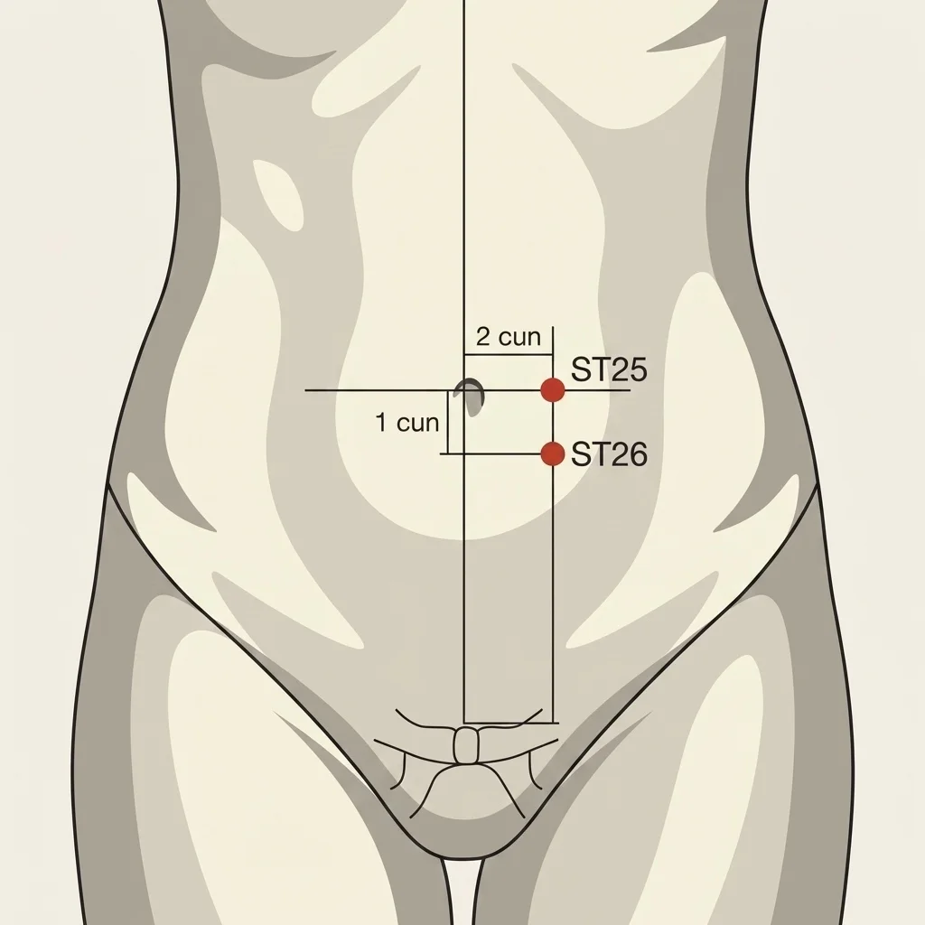
Located lateral to the umbilicus, over the rectus abdominis belly.
When to Seek Medical Help
Frequently Asked Questions About Abdominal Diastasis
Abdominal diastasis is separation of the two bellies of the rectus abdominis along the linea alba — the band of connective tissue that joins the recti at the abdominal midline. The most common cause is pregnancy, where hormones (relaxin, estrogen) combined with intra-abdominal pressure from uterine growth widen the linea alba. It can also occur in men due to abdominal obesity, excessive weightlifting, or aging.
Virtually all pregnant women have some degree of rectus separation by the end of pregnancy — a physiologic adaptation needed to accommodate uterine growth. Most recover spontaneously in the first weeks to months after delivery. About 33% retain clinically significant diastasis (greater than 2 cm) at 12 months postpartum, according to prospective studies (Sperstad et al., 2016).
Lie on your back with knees bent and feet on the floor. Place your fingers on the midline of the abdomen, above the umbilicus. Lift your head and shoulders (like a partial crunch) and feel how many fingers fit in the separation between the two muscle "cords". Repeat at the level of the umbilicus and below it. A separation of 2 or more finger-widths suggests diastasis. Also observe whether a "ridge" or cone appears at the midline — coning, which indicates that the linea alba is not tensing adequately.
Traditional abdominal exercises (crunches and sit-ups) sharply raise intra-abdominal pressure, pushing the viscera against the linea alba. When the linea alba is widened and weakened, this pressure further stretches the separation. The crunch also predominantly recruits the rectus abdominis (superficial) without adequately activating the transversus abdominis (deep) — the muscle responsible for tensing the linea alba. These exercises can be reintroduced in advanced phases, once the linea alba demonstrates functional competence.
The transversus abdominis (TrA) is the deepest abdominal muscle — it wraps around the trunk like a corset, with horizontal fibers that, when contracting, draw the rectus abdominis muscles together and tense the linea alba. For this reason, TrA activation is the starting point of rehabilitation: when it contracts adequately, the inter-recti separation reduces and the linea alba gains functional tension. Correct activation is described as "drawing the navel inward and upward", without moving the pelvis.
Yes. Diastasis in men is often underdiagnosed and is associated with abdominal obesity (chronic intra-abdominal pressure from visceral adipose tissue widens the linea alba), weightlifting with poor technique (Valsalva maneuver without TrA activation), and aging. Assessment and rehabilitation follow the same principles as postpartum — TrA activation, progressive exercises, and intra-abdominal pressure management.
Acupuncture can serve as complementary therapy, modulating the frequently associated low back pain and supporting abdominal tone. Electroacupuncture at points lateral to the linea alba (such as ST-25) can stimulate the musculature without compromising the thinned region. Safety is paramount — abdominal needling in patients with diastasis requires controlled depth; when performed by a medical acupuncturist familiar with the anatomic particularity, the procedure gains in precision and safety.
Surgery (abdominoplasty with rectus plication) is considered after 6-12 months of well-conducted rehabilitation without satisfactory improvement, especially when separation exceeds 3-4 cm, an associated hernia is present (umbilical or epigastric), or the functional and aesthetic impact significantly compromises quality of life. The decision should be evaluated by a specialist physician, since many cases respond well to conservative treatment when the exercise program is adequately supervised.
Related Reading
Deepen your knowledge with related articles