What Is Elbow Arthropathy?
Elbow arthropathy (elbow osteoarthritis) is the progressive degeneration of the articular cartilage of the elbow, accompanied by osteophyte formation (bony spurs), subchondral sclerosis, and frequently by intra-articular loose bodies. Unlike other joints, primary osteoarthritis of the elbow is relatively uncommon.
The elbow is a compound joint formed by three articulations within a single capsule: the humeroulnar (the main one for flexion/extension), the humeroradial (radiocapitellar), and the proximal radioulnar (pronation/supination). Osteoarthritis can affect one or more of these articulations.
The primary form occurs predominantly in men with heavy manual work, while the secondary form results from prior trauma (fractures, dislocations), inflammatory disease (rheumatoid arthritis), or osteochondritis dissecans.
Joint Wear
Progressive cartilage loss that produces osteophytes, loose bodies, and limited motion.
Stiff Elbow
Loss of full extension is the earliest and most frequent finding, followed by loss of flexion.
Primary vs Secondary
Primary: manual workers, athletes. Secondary: post-trauma, rheumatoid arthritis, osteochondritis.
Epidemiology
Primary elbow osteoarthritis is less frequent than hip, knee, or hand osteoarthritis, accounting for only 2% of all osteoarthritis cases. It predominantly affects men (4:1 ratio) and the dominant arm, with peak incidence between 40 and 60 years of age.
Primary osteoarthritis is particularly prevalent in manual laborers (miners, jackhammer operators, construction workers), throwing athletes (baseball, shot put), and combat sports practitioners. Repetitive high-impact loading is the main etiologic factor.
Post-traumatic secondary osteoarthritis is more common than the primary form and may occur at any age. Intra-articular elbow fractures (especially of the humeral condyle, radial head, and olecranon) are the most frequent antecedents.
Pathophysiology
Elbow osteoarthritis follows the same degenerative process as other joints: hyaline cartilage progressively wears away, the subchondral bone sclerotizes, marginal osteophytes form, and reactive synovitis develops. However, the elbow has anatomical particularities that shape its wear pattern.

Pattern of Degeneration
In primary osteoarthritis, wear predominates at the humeroulnar articulation. Osteophytes form preferentially at the tip of the olecranon, in the olecranon fossa, on the coronoid process, and in the coronoid fossa — the impact points between the humerus and ulna at the extremes of extension and flexion.
Osteophytes in the olecranon fossa block terminal extension, while those in the coronoid fossa limit flexion. This "osteophytic impingement" mechanism explains why loss of extension is the earliest finding and why the radiocapitellar joint generally remains relatively preserved in the early stages.
Intra-articular loose bodies are frequent in elbow osteoarthritis. They originate from cartilage fragments or osteophytes that detach and remain free within the joint. They may cause episodes of mechanical joint locking — sudden locking of the elbow in a given position.
Symptoms
Elbow osteoarthritis manifests progressively. Lost motion is usually the first symptom, frequently noticed by the patient before significant pain sets in. The minimum functional arc required for daily activities is 30-130 degrees of flexion-extension and 50 degrees of pronation-supination.
Symptoms of Elbow Arthropathy
Loss of full extension
The earliest finding — difficulty fully straightening the arm, noticed when washing dishes, leaning on surfaces, or extending the arm.
Pain at the extremes of motion
Mechanical pain at the end of maximum extension and flexion, caused by osteophytes impinging against the bony fossae.
Joint crepitus
A grating or coarse clicking sensation during elbow movement, perceived by the patient and palpable by the examiner.
Episodes of mechanical locking
The elbow suddenly locks in a given position, then releases — caused by intra-articular loose bodies.
Diffuse elbow pain
Deep, diffuse pain that worsens with prolonged use and improves with rest. Different from the focal pain of epicondylitis.
Intermittent joint swelling
Mild to moderate joint effusion, especially after exertion, perceived as a diffuse "swelling" around the elbow.
Diagnosis
Elbow osteoarthritis is diagnosed from a clinical history of progressive loss of range of motion with mechanical pain, confirmed by plain radiographs in anteroposterior and lateral views.
Diagnosis of Elbow Osteoarthritis
Source: Usual clinical and radiographic findings
Clinical Findings
- Loss of terminal extension (earliest and most sensitive finding)
- Crepitus on passive elbow mobilization
- Pain at the extremes of flexion and extension
- Episodes of mechanical locking (suggestive of loose bodies)
- Intermittent joint swelling
Radiographic Findings
- Osteophytes at the tip of the olecranon and in the olecranon fossa
- Osteophytes at the coronoid process and in the coronoid fossa
- Joint space narrowing (humeroulnar and/or radiocapitellar)
- Subchondral sclerosis
- Intra-articular loose bodies (calcified)
Imaging Studies in Elbow Osteoarthritis
| Study | Indication | Main Findings |
|---|---|---|
| AP and Lateral Radiograph | First choice — confirms the diagnosis | Osteophytes, joint space narrowing, calcified loose bodies, sclerosis |
| Computed Tomography | Preoperative planning | Detailed bony anatomy, precise localization of osteophytes and loose bodies |
| Magnetic Resonance Imaging | Assessment of associated soft tissues | Chondral lesions, ligament injuries, synovial plica, ulnar neuropathy |
| Ultrasonography | Assessment of effusion and soft tissues | Joint effusion, synovial thickening, superficial loose bodies |

Differential Diagnosis
Lateral Epicondylitis
Read more- Focal pain at the lateral epicondyle
- No limitation of range of motion
- Pain on resisted wrist extension
Rheumatoid Arthritis of the Elbow
- Polyarticular and symmetric involvement
- Prolonged morning stiffness
- Exuberant synovitis with warm swelling
Osteochondritis Dissecans
- Young patients (adolescents)
- Lateral elbow pain
- Mechanical locking
Radial Tunnel Syndrome
Read more- Proximal forearm pain (3-5 cm distal to the epicondyle)
- No crepitus or loss of range of motion
- Pain on resisted supination
Olecranon Bursitis
Read more- Fluctuant posterior swelling over the olecranon
- No limitation of range of motion (except extreme flexion)
- No crepitus
Treatments
Conservative treatment is the first line for most patients with symptomatic elbow osteoarthritis. The goal is to control pain, preserve functional range of motion, and slow joint degeneration.
Conservative Treatment
Active and passive joint mobilization is fundamental to maintain range of motion. Daily active flexion-extension and pronation-supination exercises, within tolerated pain limits, prevent the progression of stiffness. Gentle terminal stretching toward the limited direction is performed with caution.
Oral or topical nonsteroidal anti-inflammatory drugs (NSAIDs) help control painful flares and reduce associated synovitis. Intra-articular corticosteroid injection may be useful for acute inflammatory flares but should be limited to 3-4 applications per year.
Modifying activity — reducing impact loading and weight-bearing with the arm extended — is essential to slow progression. Replace repetitive impact activities with lower-impact exercises.
Treatments for Elbow Osteoarthritis
| Treatment | Mechanism | Evidence | Considerations |
|---|---|---|---|
| Active joint mobilization | Maintenance of range of motion, cartilage nutrition | Strong | First line — daily range-of-motion exercises |
| NSAIDs (oral or topical) | Anti-inflammatory and analgesic | Strong | Flare control; intermittent use |
| Acupuncture and laser therapy | Analgesia, modulation of inflammation (proposed mechanisms) | Limited | Adjuvant for pain; elbow-specific evidence is scarce |
| Intra-articular injection | Potent local anti-inflammatory | Moderate | Acute flares; max 3-4 per year |
| Arthroscopy (debridement) | Removal of osteophytes and loose bodies | Moderate to strong | Mechanical locking from loose bodies, significant loss of range of motion |
| Interposition arthroplasty | Tissue interposition in the joint | Moderate | Young, active patients with advanced osteoarthritis |
| Total elbow replacement | Joint replacement | Strong | Advanced osteoarthritis in low-demand patients (> 65 years) |
Do
- Do daily active flexion-extension and pronation-supination exercises, within tolerated pain limits.
- Use oral or topical nonsteroidal anti-inflammatory drugs to control painful flares.
- Replace repetitive impact activities with lower-impact exercises.
Avoid
- Bear weight with the arm extended.
- Keep the elbow at prolonged rest, which worsens joint stiffness.
- Exceed 3-4 intra-articular corticosteroid injections per year.
- Conservative treatment is the first line for most patients with symptomatic elbow osteoarthritis.
- The goal is to control pain, preserve functional range of motion, and slow joint degeneration.
- Active and passive joint mobilization is fundamental to maintain range of motion.
Acupuncture and Laser Therapy
Acupuncture may have a role as an adjuvant conservative resource in elbow osteoarthritis. The proposed mechanisms — supported mainly by experimental studies — include analgesia from endogenous opioid release, possible cytokine-mediated modulation of synovitis, and relaxation of the periarticular musculature that contributes to stiffness.
The approach includes local points around the elbow (LI11, HT3, LU5, TE10, SI8) and distal points for segmental pain modulation. Electroacupuncture with alternating frequency of 2/100 Hz is used to potentiate analgesia by activating opioidergic and serotonergic pathways described in preclinical studies.
Low-intensity laser therapy has been studied as an adjuvant for osteoarthritis of small and medium joints. Experimental studies suggest that photobiomodulation may modulate the expression of metalloproteinases, reduce synovial edema, and improve intra-articular microcirculation; the clinical translation of these findings, however, is not yet consolidated.
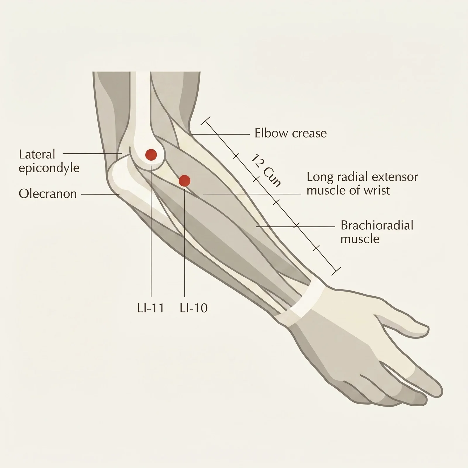
- Location
- With the elbow flexed at a right angle, at the lateral end of the cubital crease, in the depression between the end of the crease and the lateral epicondyle of the humerus, over the extensor carpi radialis longus muscle.
Prognosis
Elbow osteoarthritis is a progressive condition, but most patients maintain adequate function for daily activities with conservative treatment. Functional tolerance is better than in knee or hip osteoarthritis, since the elbow is not a weight-bearing joint.
Stepwise Approach
Initial Conservative Treatment
Daily range-of-motion exercises, intermittent NSAIDs, activity modification. Start acupuncture and laser therapy for pain control.
Intensified Conservative Treatment
If range of motion keeps declining: intra-articular injection, intensify assisted joint mobilization. Assess occupational ergonomics.
Surgical Consideration
If mechanical locking recurs or the functional arc is lost: arthroscopy to debride, remove loose bodies, and clear osteophytes.
Reconstructive Surgery
In advanced cases with severe functional impairment: interposition arthroplasty (young patients) or total replacement (low-demand elderly patients).
Myths and Facts
Myth vs. Fact
Elbow osteoarthritis only affects the elderly.
Primary elbow osteoarthritis predominantly affects men aged 40-60 with heavy manual work, and the post-traumatic form may occur at any age.
If the elbow clicks, it has osteoarthritis.
Mild, painless clicking is common and generally benign. In osteoarthritis, the crepitus is coarse, constant throughout the entire arc of motion, and comes with pain and limited range of motion.
With osteoarthritis, it is better not to move the elbow.
The opposite is true. Daily active mobilization is fundamental to maintain range of motion and cartilage nutrition. Prolonged rest worsens joint stiffness.
Elbow osteoarthritis always requires a prosthesis.
Most patients maintain adequate function with conservative treatment. Arthroscopy resolves many cases with loose bodies and osteophytic impingement. Total replacement is reserved for advanced cases in elderly patients.
When to Seek Medical Help
Frequently Asked Questions about Elbow Arthropathy
Elbow arthropathy (osteoarthritis) is the progressive degeneration of the elbow's articular cartilage, producing osteophytes, subchondral sclerosis, and frequently intra-articular loose bodies. The primary form predominantly affects men in heavy manual work or athletes in impact sports. The secondary form results from prior fractures, dislocations, rheumatoid arthritis, or osteochondritis dissecans.
The earliest symptom is loss of full extension — difficulty fully straightening the arm. As the disease progresses, pain appears at the extremes of motion (maximum extension and flexion), along with coarse crepitus during movement, episodes of mechanical locking from loose bodies, and intermittent joint swelling. The pain is mechanical: it worsens with use and improves with rest.
Diagnosis combines clinical findings (loss of range of motion, crepitus, pain at the extremes of motion) with plain radiographs in anteroposterior and lateral views. The radiograph shows osteophytes in the olecranon and coronoid fossae, joint space narrowing, and calcified loose bodies. CT is useful for surgical planning, and MRI assesses associated cartilage and ligament injuries.
Conservative treatment is the first line: daily range-of-motion exercises, intermittent NSAIDs for flares, modified impact activities, acupuncture, and laser therapy for pain control. Arthroscopy is indicated to remove loose bodies and osteophytes that cause mechanical locking or significant loss of range of motion. Total elbow replacement is reserved for advanced cases in elderly patients with low functional demand.
It may be considered as an adjuvant. Studies suggest an analgesic contribution, with proposed mechanisms that modulate pro-inflammatory cytokines intra-articularly and relax the periarticular musculature. Experimental evidence points to an effect of electroacupuncture on IL-1beta and TNF-alpha; whether this translates into a clinical chondroprotective effect is not yet a consensus. In practice, pairing acupuncture with mobilization exercises immediately after the session — taking advantage of the analgesic window — usually helps the patient regain range of motion.
Arthroscopy is indicated when loose bodies cause recurrent mechanical locking or when lost range of motion significantly compromises function despite 3-6 months of conservative treatment. Total replacement is reserved for advanced osteoarthritis in patients over 65 with low functional demand — it is not indicated for young, active patients because the implant would wear out.
Osteoarthritis is a degenerative condition without a definitive cure, but appropriate treatment can significantly slow its progression. Most patients maintain satisfactory function for daily activities with continuous conservative treatment. Unlike the knee and hip, the elbow does not bear body weight, which favors a better functional prognosis. Arthroscopy can restore range of motion by removing mechanical blocks.
Related Reading
Deepen your knowledge with related articles